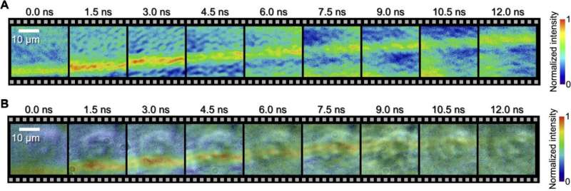
See underwater shock waves through biological cells. (A and B) STAMP movies with 9 frames spaced 1.5 ns apart showing the propagation of underwater shock waves with (B) and without (A) HeLa cells. Scale bar, 10 m. Credit: scientific progress (2023). DOI: 10.1126/sciadv.adj8608
Microscopic shock waves passing through single biological cells have been photographed thanks to a new photography technique. Nanophotography uses ultrafast electronic cameras to capture images at billionths of a second. However, image quality and exposure time are often limited.
Now, a team led by researchers at the University of Tokyo has achieved ultra-fine images taken at high speed on multiple time scales using a system they named Spectral Circuits. Spectral circuitry bridges the gap between optical imaging and traditional electronic cameras, enabling photography to be performed at ultra-fast speeds, with less blur and greater precision. This technology has potential applications in science, medicine and industry.
In photography, timing is of the essence, and capturing images at high speeds poses special challenges. But thanks to advances in camera technology, we can now see the world like never before. Whether it’s the sweat on a race car driver’s forehead, the focus of a swooping falcon’s eye, or the latest improvements in nanosecond photography, the movement of shock waves through microscopic single cells at high speeds.
Paper published in journal scientific progress.
“To our knowledge, for the first time in history, the interaction between biological cells and shock waves has been directly observed, and it has been experimentally proven that shock waves propagate faster inside the cell than outside the cell,” said Precision, University of Tokyo Takao Saiki, a doctoral student in the Department of Engineering, explains.
“In addition, our method allows us to demonstrate high-speed photography over a wide time range, including picoseconds (trillionths of a second), nanoseconds (billionths of a second), and milliseconds (thousandths of a second). seconds) ) time scale.”
Capturing clear images of cells without affecting their structure or causing damage is very challenging. To capture images safely, the researchers developed a sophisticated optical circuit that uses light instead of electricity, which they named a spectroscopic circuit. Using spectroscopic circuitry, they created non-destructive laser pulses and programmed them to fire at different times. By combining this technique with an existing single-shot optical imaging technique called sequential timed all-optical mapping photography (STAMP), they were able to capture a series of images that were clearer and less blurry than before.
The team used the same technique to study the effects of laser ablation on glass. Laser ablation can be used to precisely remove solid materials from surfaces and is used in industry and medicine. The researchers focused ultrashort laser pulses of just 35 femtoseconds (one femtosecond equals one trillionth of a second) onto a glass plate. Using spectroscopic circuits, they observed the impact of the laser, the resulting shock wave, and its effect on the glass in picoseconds, nanoseconds and milliseconds.
-
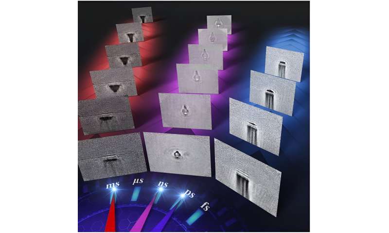
Laser ablation images captured using ultra-wide time range high-speed cameras: Using this new imaging technique, researchers can see propagating shock waves and plasma and multiple time scales (about 10,100 picoseconds, about 110 nanoseconds) of laser ablation progress of the shot processing, approximately 1100 ms). Image source: 2023 Saiki et al/CC BY NC
-
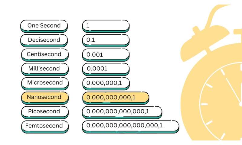
Less than a second: Picoseconds are the typical rate used in ultrafast optical imaging, while high-speed electronic cameras can capture images at millisecond and microsecond rates. The research team’s system of spectral circuits bridges the gap between these techniques, allowing us to see what is happening between these timeframes. Image source: 2023 Nicola Burghall / CC BY
“We can see the interactions between different physical processes that occur over time and how they are formed,” said Keiichi Nakagawa, associate professor in the Department of Bioengineering and Precision Engineering at the University of Tokyo. “Our technology allows us to Observing and analyzing such ultrafast processes provides the opportunity to reveal useful but unknown high-speed phenomena.
“Next, we are planning to use our imaging technique to visualize how cells interact with acoustic waves, like those used in ultrasound and shock wave therapy. By doing this, we aim to understand the primary physical processes that activate subsequent therapeutic effects in the human body.” The team also hopes to use spectroscopic circuits to improve laser processing techniques by identifying physical parameters to enable faster, more precise, more consistent and more cost-effective manufacturing.
“We have always been fascinated by the power of visualization to understand complex phenomena. The opportunity to reveal and show previously hidden parts of the world really attracted us to this field,” Nakagawa said. “We expect to make broad contributions in various fields including biomedicine, manufacturing, materials, environment and energy.”
More information:
Takao Saiki et al., Single-shot optical imaging with spectral circuit bridging time scales in high-speed photography, scientific progress (2023). DOI: 10.1126/sciadv.adj8608
Provided by the University of Tokyo
citation: Shock waves traveling through a single cell captured with improved nanosecond imaging technique (2023, December 21), Retrieved December 21, 2023, from https://phys.org/news/2023-12-cell-nanosecond -imaging-technology.html
This document is protected by copyright. No part may be reproduced without written permission except in the interests of fair dealing for private study or research purposes. Content is for reference only.
#improved #nanosecond #imaging #technique #film #shock #waves #passing #single #cell
Image Source : phys.org