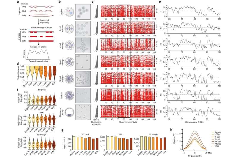
RT emerges gradually during preimplantation development in mice. AOverview of single-cell Repli-seq for generating RT profiles from single cells in mouse preimplantation embryos based on copy number variation. Second, schematic diagram of embryo sampling and corresponding images of isolated blastomeres at each stage. The number of independent blastomere sets for each stage with similar results is as follows: zygote (3), 2-cell (4), 4-cell (3), 8-cell (3), 16-cell (3) , Morula (2), ICM (4). Scale bar, 50m. C, single-cell heat map indicating replication status according to binarized copy number during preimplantation embryogenesis (red, replicated; gray, not replicated). Cells are ranked by the percentage of their genome replicated (replication fraction), which indicates progression into S phase and is plotted as the bar graph on the left. d, variation score during embryonic development; score is 1 when 50% of cells replicate the genome bin; score is 0 when all cells replicate (100%) or do not replicate (0%). Each violin plot shows the distribution of scores across all genome bins. eRT map of preimplantation embryos in a representative region on chromosome 2, represented by a black rectangle in the figure C. The black line represents the RT profile calculated as the average of overlapping intervals defined by the whole-genome copy score. F,Gsize(F) and numbers (GDuring preimplantation development, replication is characterized by RT peaks (also known as initiation zones) and RT troughs (also known as termination zones). Boxplots show the median and interquartile range (IQR), and whiskers depict the lowest and highest values within 1.5IQR. bp, base pair. H, the relative RT values are concentrated at the RT peak during embryonic development compared with adjacent regions. Note that the curves for the 2-cell and 4-cell stages overlap considerably and to some extent overlap with the curve for the zygote. Credit: nature (2023). DOI: 10.1038/s41586-023-06872-1
The complex process of copying genetic information, called DNA replication, is at the heart of the transmission of life from one cell to another and from one organism to the next. This is accomplished not only by copying genetic information; A carefully planned series of molecular events must also occur at the right time.
Scientists in collaboration with Professor Maria-Elena Torres-Padilla from Helmholtz München recently discovered a fascinating aspect of this process, known as “replication timing” (RT), and the peculiarities of the beginning of life .The new results are now published in nature.
The process of DNA replication timing (RT) refers to the specific moments at which different regions of our genetic code are copied. Researchers at the Helmholtz Institute for Epigenetics and Stem Cell Research in Munich used a technology called “Repli-seq” to delve into the close relationship between RT and cell adaptability (cell plasticity).
Interestingly, they also discovered a new relationship between RT and how genes fold into three-dimensional structures within the nucleus.
Starting from the earliest stages of the embryo, the fertilized egg (the earliest stage of an organism’s life), the researchers created RT maps from the single-cell stage to the stage when the embryo implants in the mother’s uterus, called the blastocyst. An unexpected finding was that RT in single-cell embryos is not very ordered, suggesting that genome replication is very flexible in these early cells.
However, after the 4-cell stage, RT becomes more defined. A progressive process is taking place, reflecting the gradual acquisition of modifications of DNA and associated proteins, so-called chromatin marks, that indicate a gene’s activity and importance in cellular function.
Maria-Elena Torres-Padilla, corresponding author of the study, further explains: “This is noteworthy because it tells us that these early embryonic cells have a very ‘plastic’ genome replication program. Because these early cells are totipotent, they can create “Every cell in our body. We think what we discovered in this study is one of the reasons why these cells are so good at generating the entire body. “
New discoveries about DNA replication could become tools for reprogramming cells. Dr. Tsunetoshi Nakatani, first author of the study, added: “We can envision changing cell identity by changing the RT procedure to a more flexible one.”
The results further suggest that RNA polymerase, commonly known as the enzyme responsible for reading the genetic code and transcribing it into RNA, helps determine the precise RT program, providing some clues as to how such a program might be manipulated in the future.
The team found that the three-dimensional structure of the genome was formed first, and the RT program was established from there. This is an exciting finding because it suggests that how our genome fits into the three-dimensional space of the nucleus affects the flexibility of the RT program.
In summary, the timing of DNA replication is a fascinating piece of the puzzle in the grand narrative of life. It demonstrates how the precision of gene replication is closely related to the ability of early embryonic cells to generate other cell types in our bodies. As researchers continue to explore these connections, we are gaining a deeper understanding of the nature of life’s transmission, cell to cell, organism to organism, and the factors that enable cells to create new bodies.
More information:
Maria-Elena Torres-Padilla, The emergence of replication timing during early mammalian development, nature (2023). DOI: 10.1038/s41586-023-06872-1
Courtesy of the German Research Center Helmholtz Association
citation: Team discovers relationship between DNA replication time and how genes fold into 3D structures within cell nuclei (2023, December 20), retrieved from https://phys.org/news/2023- on December 21, 2023 12-team-relationship-dna-replication-genes.html
This document is protected by copyright. No part may be reproduced without written permission except in the interests of fair dealing for private study or research purposes. Content is for reference only.
#Team #discovers #relationship #DNA #replication #time #genes #fold #structures #cell #nucleus
Image Source : phys.org